FDA records indicate that there are no current recalls for this drug.
Are you a medical professional?
Trending Topics
Datscan Recall
Get an alert when a recall is issued.
Questions & Answers
Side Effects & Adverse Reactions
There is currently no warning information available for this product. We apologize for any inconvenience.
Legal Issues
There is currently no legal information available for this drug.
FDA Safety Alerts
There are currently no FDA safety alerts available for this drug.
Manufacturer Warnings
There is currently no manufacturer warning information available for this drug.
FDA Labeling Changes
There are currently no FDA labeling changes available for this drug.
Uses
DaTscan is a radiopharmaceutical indicated for striatal dopamine transporter visualization using single photon emission computed tomography (SPECT) brain imaging to assist in the evaluation of adult patients with suspected Parkinsonian syndromes (PS). In these patients, DaTscan may be used to help differentiate essential tremor from tremor due to PS (idiopathic Parkinson's disease, multiple system atrophy and progressive supranuclear palsy). DaTscan is an adjunct to other diagnostic evaluations.
History
There is currently no drug history available for this drug.
Other Information
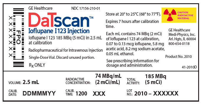
DaTscan [Ioflupane I 123 Injection] is a sterile, pyrogen-free radiopharmaceutical for intravenous injection. The clear and colorless solution is supplied in single-use vials in which each milliliter contains 0.07 to 0.13 µg ioflupane, 74 MBq (2 mCi) of iodine 123 (as ioflupane I 123) at calibration time, 5.7 mg acetic acid, 7.8 mg sodium acetate and 0.05 mL (5%) ethanol. The pH of the solution is between 4.2 and 5.2. Ioflupane I 123 has the following structural formula:

Iodine 123 is a cyclotron-produced radionuclide that decays to 123Te by electron capture and has a physical half-life of 13.2 hours. The photon that is useful for detection and imaging studies is listed in Table 2.
| Radiation | Energy Level (keV) | Abundance (%) |
|---|---|---|
| Gamma | 159 | 83 |
The specific gamma-ray constant for iodine 123 is 1.6 R/mCi-hr at 1 cm. The first half-value thickness of lead (Pb) for iodine 123 is 0.04 cm. The relative transmission of radiation emitted by the radionuclide that results from interposition of various thicknesses of Pb is shown in Table 3 (e.g., the use of 2.16 cm Pb will decrease the external radiation exposure by a factor of about 1,000).
| Shield Thickness cm of lead (Pb) | Reduction in In-air Collision Kerma |
|---|---|
|
|
| 0.04 | 0.5 |
| 0.13 | 10-1 |
| 0.77 | 10-2 |
| 2.16 | 10-3 |
| 3.67 | 10-4 |
Sources
Datscan Manufacturers
-
Medi-physics Inc. Dba Ge Healthcare.
![Datscan (Ioflupane I-123 And Iodine) Injection, Solution [Medi-physics Inc. Dba Ge Healthcare.]](/wp-content/themes/bootstrap/assets/img/loading2.gif)
Datscan | Medi-physics Inc. Dba Ge Healthcare.
![Datscan (Ioflupane I-123 And Iodine) Injection, Solution [Medi-physics Inc. Dba Ge Healthcare.] Datscan (Ioflupane I-123 And Iodine) Injection, Solution [Medi-physics Inc. Dba Ge Healthcare.]](/wp-content/themes/bootstrap/assets/img/loading2.gif)
2.1 Radiation SafetyDaTscan emits radiation and must be handled with safety measures to minimize radiation exposure to clinical personnel and patients. Radiopharmaceuticals should be used by or under the control of physicians who are qualified by specific training and experienced in the safe use and handling of radionuclides, and whose experience and training have been approved by the appropriate government agency authorized to license the use of radionuclides. DaTscan dosing is based upon the radioactivity determined using a suitably calibrated instrument immediately prior to administration.
To minimize radiation dose to the bladder, encourage hydration prior to and following DaTscan administration in order to permit frequent voiding. Encourage the patient to void frequently for the first 48 hours following DaTscan administration [see Dosage and Administration (2.5)].
2.2 Thyroid Blockade Before DaTscan InjectionBefore administration of DaTscan, administer Potassium Iodide Oral Solution or Lugol's Solution (equivalent to 100 mg iodide) or potassium perchlorate (400 mg) to block uptake of iodine 123 by the patient's thyroid. Administer the blocking agent at least one hour before the dose of DaTscan [see Warnings and Precautions (5.2)].
2.3 Preparation and AdministrationUse aseptic procedures and radiation shielding during preparation and administration. Inspect the DaTscan vial prior to administration and do not use it if the vial contains particulate matter or discoloration [see Description (11)]. Administer DaTscan as a slow intravenous injection (administered over a period of not less than 15 to 20 seconds) via an arm vein.
2.4 Recommended DoseThe recommended dose is 111 to 185 MBq (3 to 5 mCi) administered intravenously [see Clinical Studies (14)].
2.5 Radiation DosimetryThe estimated radiation absorbed doses to an average adult from intravenous injection of DaTscan are shown in Table 1. The values are calculated assuming urinary bladder emptying at 4.8-hour intervals and appropriate thyroid blocking (iodine 123 is a known Auger electron emitter).
Table 1 Estimated Radiation Absorbed Doses from DaTscan ORGAN / TISSUE ABSORBED DOSE PER UNIT ADMINISTERED ACTIVITY
(µGy / MBq) *The absorbed dose to the colon wall is the mass-weighted sum of the absorbed doses to the upper and lower large intestine walls, D Colon = 0.57 DULI + 0.43 DLLI [Publication 80 of the ICRP (International Commission on Radiological Protection); Annals of the ICRP 28 (3). Oxford: Pergamon Press; 1998]
Adrenals 12.9 Brain 17.8 Striata 230.0 Breasts 7.8 Esophagus 10.0 Gallbladder Wall 26.4 GI Tract Stomach Wall 11.2 Small Intestine Wall 21.2 Colon Wall * 39.8 Upper Large Intestine Wall 38.1 Lower Large Intestine Wall 42.0 Heart Wall 12.9 Kidneys 10.9 Liver 27.9 Lungs 41.2 Muscle 9.4 Osteogenic Cells 28.2 Ovaries 16.8 Pancreas 13.0 Red Marrow 9.2 Skin 6.0 Spleen 10.4 Testes 8.5 Thymus 10.0 Thyroid 9.0 Urinary Bladder Wall 53.1 Uterus 16.1 Total Body 11.3 EFFECTIVE DOSE PER UNIT ADMINISTERED ACTIVITY
(µSv / MBq) 21.3The Effective Dose resulting from a DaTscan administration with an administered activity of 185 MBq (5 mCi) is 3.94 mSv in an adult.
2.6 Imaging GuidelinesBegin SPECT imaging 3 to 6 hours following DaTscan administration. Acquire images using a gamma camera fitted with high-resolution collimators and set to a photopeak of 159 keV with a ± 10% energy window. Angular sampling should be not less than 120 views over 360 degrees. Position the subject supine with the head on an off-the-table headrest, a flexible head restraint such as a strip of tape across the chin or forehead may be used to help avoid movement, and set a circular orbit for the detector heads with the radius as small as possible (typically 11 to 15 cm).
Experimental studies with a striatal phantom suggest that optimal images are obtained with matrix size and zoom factors selected to give a pixel size of 3.5 to 4.5 mm. Collect a minimum of 1.5 million counts for optimal images.
2.7 Image InterpretationDaTscan images are interpreted visually, based upon the appearance of the striata. Reconstructed pixel size should be between 3.5 and 4.5 mm with slices 1 pixel thick. Optimum presentation of the reconstructed images for visual interpretation is transaxial slices parallel to the anterior commissure-posterior commissure (AC-PC) line. Determination of whether an image is normal or abnormal is made by assessing the extent (as indicated by shape) and intensity of the striatal signal. Image interpretation does not involve integration of the striatal image appearance with clinical signs and/or symptoms.
Normal :
In transaxial images, normal images are characterized by two symmetric comma- or crescent-shaped focal regions of activity mirrored about the median plane. Striatal activity is distinct, relative to surrounding brain tissue (Figure 1).
Abnormal :
Abnormal DaTscan images fall into at least one of the following three categories (all are considered abnormal).
Activity is asymmetric, e.g. activity in the region of the putamen of one hemisphere is absent or greatly reduced with respect to the other. Activity is still visible in the caudate nuclei of both hemispheres resulting in a comma or crescent shape in one and a circular or oval focus in the other. There may be reduced activity between at least one striatum and surrounding tissues (Figure 2). Activity is absent in the putamen of both hemispheres and confined to the caudate nuclei. Activity is relatively symmetric and forms two roughly circular or oval foci. Activity of one or both is generally reduced (Figure 3). Activity is absent in the putamen of both hemispheres and greatly reduced in one or both caudate nuclei. Activity of the striata with respect to the background is reduced (Figure 4).
Login To Your Free Account


